![]()
We have two major goals: defining the biological effects of the aging acetylome and non-invasive molecular imaging in the living eye for early disease detection and personalized medicine.
We have organized around two central, complementary themes: one focused on unraveling the complex story of molecular changes to the genetic material of aging cells, and the other on developing novel early-diagnostic and therapeutic technologies to identify and arrest disease processes early in their courses.
The Aging Acetylome
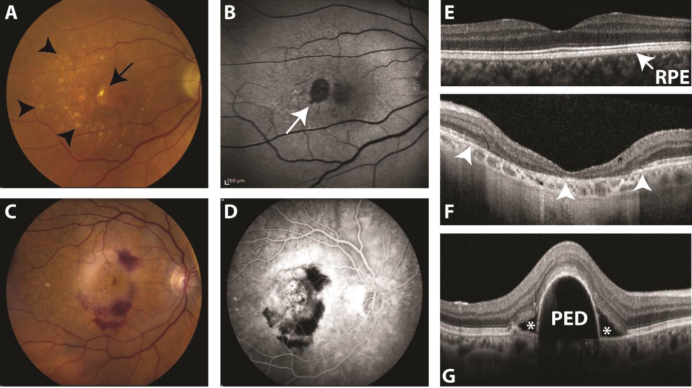
Age-related macular degeneration (AMD) is the most common cause of irreversible blindness in developed countries. Previous reports have identified increased eotaxin expression in retinal pigment epithelium (RPE) in both dry and neovascular AMD.
New insights into epigenetic derangements and chromatin remodeling have garnered great interest as a potent biologic regulatory system. Histone deacetylases (HDAC) are part of this epigenetic regulatory system and serve as a unique control of the chromatin remodeling process through regulation of post-translational modification of acetylation on histone and other non-histone proteins.
HDAC inhibitors are reported to suppress neovascularization in various mouse models and are being employed off-label in the treatment various retinal dystrophies and AMD. However, the effects of these therapies on chemokine expression in the chorioretinal microenvironment remain very unclear.
Ongoing work in the lab is utilizing the latest techniques in molecular biology to quantitatively assess expression levels and functional effects of various HDAC classes in dry AMD compared to age-matched controls as well as in cell culture and animal models. Our research aims to open translational avenues to advance dry AMD therapeutics while exploring the role of this epigenetic phenomenon is the aging retina. These studies will be greatly beneficial to the field of ophthalmology and visual science where our understanding of postnatal epigenetics and its role in aging diseases of the eye remain unclear.
Non-Invasive Molecular Imaging in the Living Eye
The ophthalmoscope, an ingenious handheld device to illuminate and visualize the eye, was introduced in 1850. In the following years, this valuable tool was widely sold across Europe resulting in a massive explosion of information pertaining to eye disease and pathogeneses of vision loss.
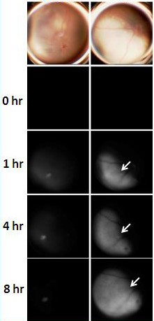
Ophthalmic fundus photography finally arrived in the early 20th century, but it was another 50 years until another major advanced occurred with the discovery of fluorescein angiography, a technique developed by two American medical students. The novel procedure utilized a non-toxic green fluorescent dye delivered intravenously to delineate the retinal and choroidal vasculature. Indocyanine green was later introduced for improved choroidal imaging, as its spectral characteristics in the near-infrared region allowed for enhanced signal through the overlying RPE.
Today, ophthalmologists and retinal specialists around the world still rely on these angiography techniques that were developed over 50 years ago. Yet, there are now a multitude of known fundamental molecular mechanisms that drive a number of vision and life threatening eye diseases.
In my lab, we are rigorously investigating new approaches to safe and feasible ocular bio-imaging in specific vision and life threatening eye diseases using the latest advancements in fluorescence technology and biomedical engineering.
